Semen Sample Under the Microscope
作者:
Karim Abdelmomen
最近上传:
10 年前
许可:
Creative Commons CC BY 4.0
摘要:
The objective of thiss experiment is to examine how a semen sample will look like under a microscope and to scale it and take the its measurements.

The objective of thiss experiment is to examine how a semen sample will look like under a microscope and to scale it and take the its measurements.

\documentclass{MNR}
%\setcounter{page}{}
%\journaltitle{}
%\volume{}
%\pubyear{}
%\doi{}
%\articlenumber{}
%\articletype{}


\articletitle{} % To break lines in long titles use \\
%%%%%%%%%%%%%%%%%%%%%%%%%%%%%%%%%%%%%%%%%%%%%%%%%%%%%%%%
%%%
%%%  Main document.
%%%
%%%  To span floating objects (wide images, tables, etc.)
%%%  over twocolumn layout, use figure*
%%%  or table* environments.
%%%
%%%  Examples:
%%%  \begin{figure*}	 \begin{table*}
%%%   ...				  ...
%%%  \end{figure*}		 \end{table*}
%%%
%%%%%%%%%%%%%%%%%%%%%%%%%%%%%%%%%%%%%%%%%%%%%%%%%%%%%%%%
\begin{document}
\maketitle
\section*{Preparation of the Semen Sample Slide}
The sample was collected from a male of the age ranging between 20-25 The donor's ejaculation was taken and immediately placed in an incubator set to the temperature of 37 degrees Celsius. The sample stayed their for a period of 30 minutes for it to liquefy completely.
A 3 ml dropper was then used to take a drop of the sample and placed on a microscope slide and placed under the microscope fitted with a lens of 50x magnification, Mitutoyo microscope.

Another drop was taken to prepare another microscope slide to be placed under another microscope to measure the scale, Axio microscope.
These experiments were conducted in the MNR lab in the German University in Cairo, GUC.

\section*{Results}

\subsection*{Mitutoyo Results}
The Mitutoyo showed a living sample of the semen and showed the sperms moving easily in the semen. There was a large number of sperms moving around the semen. There were also some deformed sperms and a few were dead.

\begin{figure}[t]
\centering
\includegraphics[width=2.5in]{2.jpg}
\caption{HERAtherm Incubator}\label{fig01:System}
\end{figure}

\subsection*{Axio Results}
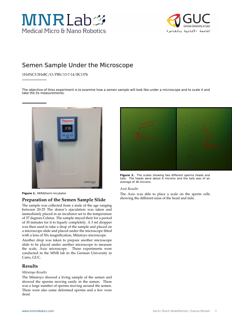
The Axio was able to place a scale on the sperm cells showing the different sizes of the head and tails.

\begin{figure}[t]
\centering
\includegraphics[width=4in]{Collage.jpg}
\caption{The scales showing two different sperms heads and tails. The heads were about 6 microns and the tails was of an average of 36 microns.}\label{fig01:System}
\end{figure}

\end{document}